
3D/4DUltrasound—Real-TimeImagingoftheFetus
Written & medically reviewed by Dr. Shaivalini Kamarapu
MBBS · MS (Obstetrics & Gynaecology) · Fellowship in Reproductive Medicine · 20+ years of clinical experience
0+
Years of Experience
0+
Successful Surgeries
0+
Happy Families
3D/4Dultrasound—real-timeimagingofthefetus
At AMVI Hospital, we provide 3D and 4D ultrasound scanning facilities. This allows our doctors to inspect the fetus and the uterus, including the amount of amniotic fluid, and to observe for any abnormalities in the growing fetus. These scans are recommended for high-risk pregnancies to monitor the growing fetus and the mother's uterus.
At AMVI Hospital, we have a team of experts to help you throughout your pregnancy. We have the medical infrastructure and utilities to provide all-round care and support to patients, with an expert team of doctors to organize ultrasound scanning appointments and provide proper care from the beginning of pregnancy through to childbirth.
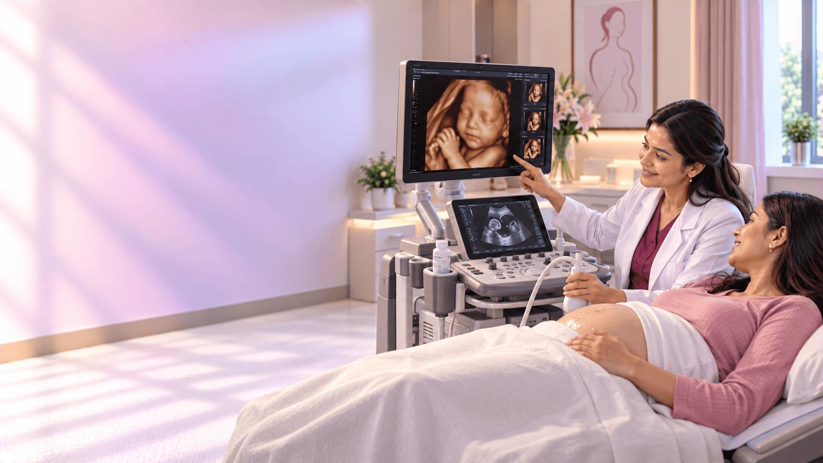
Ultrasound scans, including 3D and 4D, are more like a real-time photograph of your baby. With 3D scanning, many pictures of the child are taken in 2D and then merged to create a 3D image effect. With 4D scanning, pictures are made in real time, so you can see what your child is doing in the womb at that moment — such as moving their legs and arms, or opening their eyes.
Ultrasound scans are an essential tool to examine the health and internal organs of a growing fetus. They help gynaecologists identify complications early, so they can be treated at the earliest opportunity. 3D/4D scans performed throughout pregnancy help detect anomalies such as a cleft lip or underdeveloped limbs, help track spinal problems and other congenital conditions, and help monitor amniotic fluid levels.
Reasonstogetultrasoundsduringpregnancy
Confirming your estimated due date
Looking at your baby's heartbeat
Making sure the pregnancy isn't ectopic (i.e. in the fallopian tubes) and is in the uterus
Confirming the number of babies in utero
Making sure baby is developing properly and at the appropriate pace
Checking and measuring baby's major organs
Measuring the size of your baby
Checking amniotic fluid levels
Ruling out certain birth defects
Determining baby's sex
Giving parents a look at baby and reassurance that all is going as it should
Why3Dand4Dsonogramsareperformedduringpregnancy
Medical practitioners use 2D and Doppler ultrasounds in uncomplicated pregnancies to examine the fetus, assess amniotic fluid, and look for birth defects, among other reasons.
3D and 4D ultrasounds are performed only to closely examine suspected fetal anomalies, such as cleft lip and spinal cord issues, or to monitor something specific. In other words, 3D and 4D scans are usually not part of routine prenatal exams.
What'sthedifferencebetween2D,Doppler,3Dand4Dultrasounds?
2D Ultrasound
A wand (transducer) placed on your belly or into your vagina sends sound waves through your body. The waves bounce off internal organs and fluids, and a computer converts these echoes into a two-dimensional cross-sectional image of the fetus on a screen.
Doppler
Your practitioner uses a hand-held ultrasound device, with the help of a special jelly on your belly, to amplify the sound of the fetal heartbeat.
3D Ultrasound
Multiple two-dimensional images are taken at various angles and then pieced together to form a three-dimensional rendering — instead of just a profile view, you can see the whole surface of your baby's face, looking more like a regular photo.
4D Ultrasound
Similar to a 3D ultrasound, but the image shows movement like a video would — so you'd see your baby doing things in real time, like opening and closing their eyes or sucking their thumb.
Benefits of 3D scan
A better view of foetal heart structures, producing images that cannot be achieved through 2D models
Helps diagnose neural defects and foetal musculoskeletal issues
Helps diagnose foetal face defects such as a cleft lip
Takes less time to get the standard view of the plane
The recorded volume data can be kept on record for better diagnosis and expert advice
Easy to study and understand
Benefits of 4D scan
Increases parenting connection with the child
Shorter time for foetal heart screening and diagnosis
The recorded data can be kept on record for expert review
Shares the advantages of 3D ultrasound, including examining the foetal heartbeat, placental localization, and assessing foetal wellbeing and growth